페이지 위치
Bone Density Test
- Exam. Time :20 mins
- Price :45,000 won
Measuring bone density is valuable for predicting the risk of fractures in any area, but measuring a specific area is the best method to predict fractures in that area.
For example, to forecast the risk of fracturing a femur, it is best to measure the bone density of the femur.
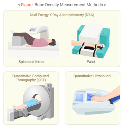
For the bone density measurement method for clinical use, dual energy x-ray absorptiometry (DXA) is most widely used, and there are various other methods such as quantitative computed tomography (QCT), ultrasound, etc.
When determining bone density, T-values and Z-values are mainly used, rather than the absolute values measured.
T-values are the standard deviation compared to the average bone density of the young adults in the same gender group, which mean the difference from healthy adults.
On the other hand, Z-values represent a difference from the average bone density of the same age group.
As to postmenopausal women and men over 50, osteoporosis is diagnosed according to T-values, and infants, adolescents, premenopausal women and men under the age of 50 are judged by Z-values.
If the T-value is less than -2.5, osteoporosis is diagnosed, and -1.0 to -2.5 will be determined as osteopenia. If the Z-value is less than -2.0, it is defined as ‘below the expected range for age’ and the possibility of secondary osteoporosis should be considered.

1. Hanaro’s Bone Density Measurement Method
Dual Energy X-Ray Absorptiometry (DXA)
There are methods for measuring the central bones such ash backbone and thighbone, and for measuring the terminal bones such as upper limbs, feet and hands bones.
The World Health Organization's diagnostic method is based on measurements of the spine and femur, and if these two areas cannot be measured, it is recommended to measure the upper limb (the wrist).
Recommended Target
Currently, the International Bone Density Institute recommend the bone density measurements for:
- Women over 65 and men over 70
- Postmenopausal women and men aged 50-69 with risk factors
- Postmenopausal women who are light in weight, are taking high-risk medicines and have experienced fractures
- Adults who have experienced fractures after the age of 50
- Those who have a disease or are taking drugs that can cause osteoporosis
- Postmenopausal women who have stopped female hormonal treatment
- To determine the effectiveness of osteoporosis medicine treatment

